
THE CONTENTS OF THE FOLLOWING PAGE ARE FOR INFORMATIONAL PURPOSES ONLY AND ARE NOT A SUBSTITUTE FOR DIAGNOSIS, MEDICAL ADVICE OR RECOMMENDATIONS.
PLEASE CONTACT YOUR TREATMENT TEAM FOR MORE INFORMATION.
Intraoperative neurophysiological monitoring (IONM) enables nerve-sparing treatment of scoliosis in spine surgery
 Back
Back
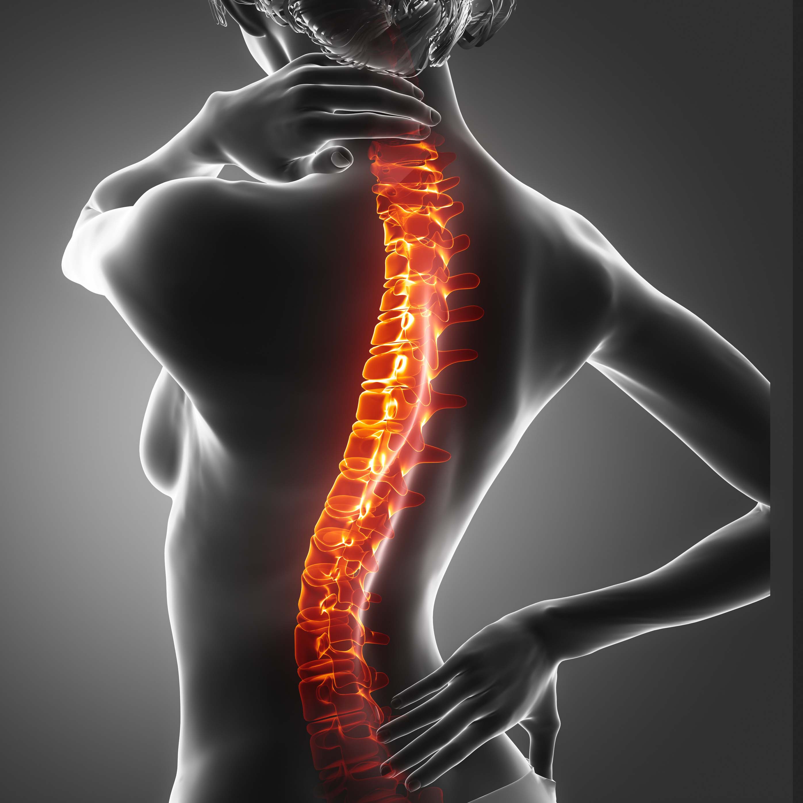
Scoliosis – deformation of the spine
Scoliosis is a deformation of the spine that is accompanied by a twisting of the vertebral bodies and bending of the spine, usually in a lateral direction. A prerequisite for the diagnosis of scoliosis is a curvature of the spine of at least 10 degrees (Cobb angle). Different degrees of severity can be distinguished on the basis of the angle of curvature.[1]
Scoliosis is usually treated conservatively in the first step (physiotherapy, corset, etc.). If this is not (or is no longer) sufficient, the surgical treatment of scoliosis is an effective but complex intervention and depends on different factors such as age, cause and severity of the scoliosis. The earlier the diagnosis is made, the greater the likelihood of non-surgical treatment.
In advanced scoliosis, from about 45°, conservative methods are usually exhausted, so that surgical intervention must be considered.[2]
Monitoring during scoliosis surgery protects against spinal cord injuries
Surgical interventions on the spine are amongst the most demanding procedures in orthopaedic surgery in terms of technique and effort. The spinal cord with its important sensory and motor pathways and spinal nerves runs within the spinal canal protected by the vertebral bones. During surgical interventions of the spine, the spinal cord can therefore be damaged, for example, by pressure, tension, bruising or, rarely, cutting. With the help of IONM, the nerve fibres of the spinal cord are continuously monitored and the smallest changes in motor and sensory functions can be detected at an early stage.[3]
Surgical treatment of scoliosis by means of spinal fusion
One method of treating scoliosis is sectional spinal fusion using screws (pedicle screws). Here, the fixation and stabilisation of the corresponding spinal segment is done by a screw-rod system. During spinal fusion, exact positioning of the screws is crucial for the success of the treatment. In addition to nerve monitoring, IONM also serves as an instrument for correct positioning of the screws. By using IONM, the surgeons can quickly and reliably check the position of the screws and prevent possible misplacement and resulting nerve damage. When the correction is done and the spine is repositioned by the rod system, the spinal cord, spinal roots and the vessels necessary for the blood flow in the spinal cord can be stretched and damaged. Continuous IONM can help find the right amount of correction during this critical stage of the procedure.
IONM procedure for nerve monitoring/pedicle screw placement
>> Placement of needle electrodes on arms, legs and head for stimulation and recording
>> These muscle movements and the sensory signals are recorded via the placed electrodest
>> Nerve monitoring is carried out by means of small current pulses that either trigger a muscle movement (motor signal) or produce a sensation in the brain region responsible for it (sensory signal)
>> Evaluation and representation of the response signals by the monitoring device
If the signal remains constant during the entire surgery, consequential damage can be almost completely ruled out. If the signal changes, e.g. due to bruising of the spinal canal during correction, the surgical strategy can be adjusted, which in most cases leads to a recovery of the signals.
In addition to the treatment of scoliosis, another field of application of intraoperative neuromonitoring is the surgical removal of colorectal cancer.
References
1. Kaye A. D. et al. Principles of Neurophysiological Assessment, Mapping and Monitoring: Clear and comprehensive coverage of clinically relevant anatomy and physiology (2014).
2. Kaye A. D. et al. Principles of Neurophysiological Assessment, Mapping and Monitoring: Clear and comprehensive coverage of clinically relevant anatomy and physiology (2014).
3. Ulkatan S. et al. Monitoring of Scoliosis Surgery with Epidurally Recorded Motor Evoked Potentials (D Wave) Revealed False Results, Clinical Neurophysiology 117, Nr. 9 (2006).
Diseases
Navigation:
| Continue to ENT |
| Go back to the overview for patients |