
THE CONTENTS OF THE FOLLOWING PAGE ARE FOR INFORMATIONAL PURPOSES ONLY AND ARE NOT A SUBSTITUTE FOR DIAGNOSIS, MEDICAL ADVICE OR RECOMMENDATIONS.
PLEASE CONTACT YOUR TREATMENT TEAM FOR MORE INFORMATION.
Neuromonitoring during surgical treatment of narrowing of the carotid artery (carotid artery stenosis)
 Vessel
Carotid artery
A common cause of stroke is calcification and associated narrowing of the carotid artery (common carotid artery and internal carotid artery). Surgical intervention on the carotid artery can prevent occlusion of this artery and the reduced blood flow to brain vessels, preventing an impending stroke. In this context, intraoperative neuromonitoring (IONM) is an important supporting technology.
The carotid artery has an important brain-supplying function
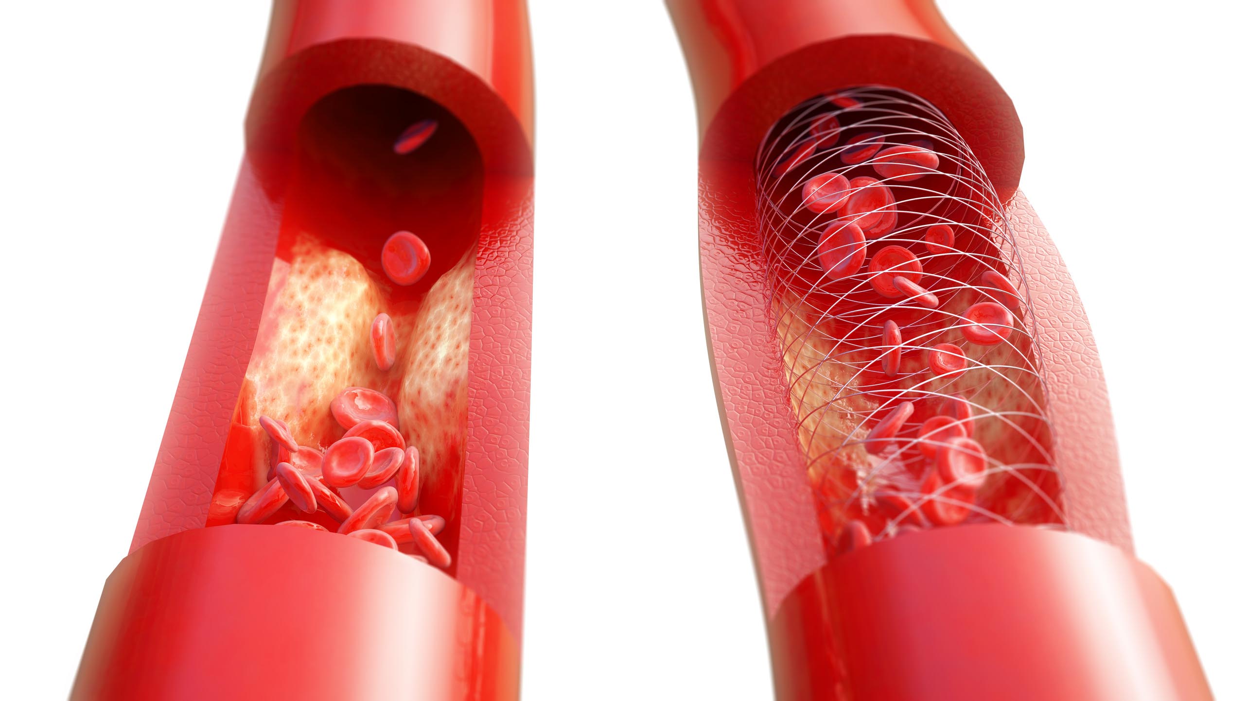
The common carotid artery is divided into an external and internal artery and supplies the brain with blood. Calcifications on the vessel wall, known as arteriosclerosis, often form at the point where the common carotid artery divides into the internal and external artery. The blood flow is disrupted and the blood supply to the brain can be affected. Blood clots that detach from the calcified vessel wall and reach the brain can also lead to an occlusion of brain vessels. The consequences can be dizziness, visual and speech disturbance, movement disorders of the arms and legs, and even a stroke.[2] If the carotid artery has a degree of narrowing of 70% or more, the risk of stroke is particularly high and treatment is recommended.[1]
Surgery on the carotid artery
During surgery, the carotid artery on the affected side is clamped and the narrowed section of the vessel is exposed through a small incision. The calcifications are removed, and both ends of the artery are sutured after cleaning. Alternatively, a vascular support (stent) can be inserted. Stents are made of either plastic or a part of the body’s own vein. Well over 25,000 of these surgeries are performed in Germany every year.[1]
Use of neuromonitoring during surgery on the carotid artery
Neuromonitoring is used to monitor somatosensory evoked potentials (SEP). By measuring the SEP signals during a surgical intervention on the carotid artery, reduced blood flow due to the clamping of the brain vessels can be indicated at an early stage.[3] The SEP signals arise in response to the stimulation of central nerves and show their conductivity. A change in these signals can be an indication of an incipient stroke.[4]
IONM procedure during treatment of carotid artery stenosis
>> Placement of needle electrodes in the scalp and neck area to record brain activity
>> Brain activity is measured by a stimulus generated via an electrical impulse on the wrist
>> Recording of the response signals resulting from the processing of the stimulus in the brain
>> Evaluation and representation of the signals by the monitoring device
IONM is generally used to monitor the function of potentially endangered nerves during surgeries. Neuromonitoring at the carotid artery in this case indirectly monitors the blood flow to the brain by changing the SEP nerve signals. If there are significant changes, this is a sign of inadequate blood supply.
In addition to the treatment of carotid artery stenosis, intraoperative neuromonitoring can also be used in the surgical removal of a brain tumour.
References
1. Carotisstenose. Available at: https://hirnstiftung.org/carotisstenose-schlaganfall-durch-verstopfte-halsschlagadern/. (Last accessed on: 6 November 2025).
2. Deutsche Gesellschaft für Gefaesschirurgie: Carotisstenose. Available at: https://www.gefaesschirurgie.de/patienten/gefaesserkrankungen/verengung-der-halsschlagader-carotisstenose. (Last accessed on: 6 November 2025).
3. Branston, N. M., Symon, L., Crockard, H. A. & Pasztor, E. Relationship between the cortical evoked potential and local cortical blood flow following acute middle cerebral artery occlusion in the baboon. Exp. Neurol. 45, 195–208 (1974).
4. Lam, A. M., Manninen, P. H., Ferguson, G. G. & Nantau, W. Monitoring electrophysiologic function during carotid endarterectomy: a comparison of somatosensory evoked potentials and conventional electroencephalogram. Anesthesiology 75, 15–21 (1991).